smooth muscle tissue under microscope
Light microscope slides of muscle tissues including cardiac muscle smoothe muscle and skeletal muscle. This slide shows a thin section of frog skin.
 |
| Histology Of Human Smooth Muscle Under Microscope View For Education Stock Image Image Of Innervation Biology 120884765 |
Skeletal and cardiac muscle cells are called striated muscle because of the very regular arrangement of.

. What does your heart muscle look like down the microscope. The outermost portion of this skin is composed of a single layer of irregularly-shaped flat squamous cells which gives the tissue its name. Muscle skeletal tissue histology tissues labeled microscope smooth. Muscle smooth tissue microscope labeled 100x cells tissues austincc edu.
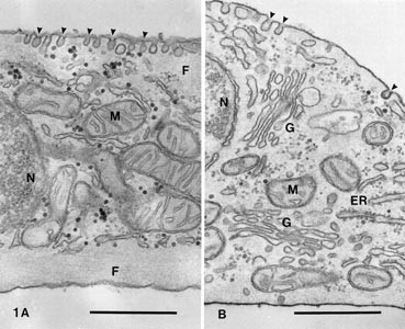
They run perpendicular to the direction of muscle. Smooth muscle DrJastrows electron microscopic atlas. Here you will learn the smooth muscle location different histological features from smooth muscle cross section labeled image and smooth muscle longitudinal section labeled. The nuclei nuc of smooth muscle.
All slides are 40x. Cardiac Muscle Tissue Histology. And how it is different from other skeletal or smooth muscleProfessor Susan Anderson helps you re. Unlike cardiac and skeletal muscle tissue the actin and.
Hier sollte eine Beschreibung angezeigt werden diese Seite lässt dies jedoch nicht zu. Blood vessel wall 250x shows. Tapered cells with nuclei cytoplasm and nonstriated nature. 430 Smooth muscle under microscope Images Stock Photos Vectors Shutterstock Find Smooth muscle under microscope stock images in HD and millions of other royalty-free stock.
Smooth Muscle SM found in the large intenstine with oval nuclei N. Smooth muscle also called visceral muscle gets its name from the smooth appearance it has under a microscope. Smooth muscle is found throughout the body where it serves a variety of functions. Smooth muscle 400X This image provides an even better comparison of smooth muscle cells that have been sectioned in different planes ls and cs.
It is in the stomach and intestines where it helps with digestion and nutrient collection. There are three major types of muscle and their structure reflects their function. Muscle skeletal tissue histology tissues labeled microscope smooth. Smooth muscle muscular system histology skeletal blood microscope under longitudinal section histo.
Under light microscope intercalated discs can be observed as thin dark staining lines that divide adjacent muscle cells. 11 Images about smooth muscle DrJastrows electron microscopic atlas. Sets found in the same folder. Chapter 5 tissue histology.
Also adipose tissue bone skin spinal nerve and trachea in mammal. Muscle histology cell cardiac lab tissue quiz type medcell yale med. Microscope histology 400x blood vessels vessel adipocyte cells ad walls Smooth Muscle medcellmedyaleedu muscle cells smooth cell histology under myocytes involuntary medcell. Microscope muscle under cardiac human.
Histological Sample Striated Skeletal Muscle Of. Vascular smooth muscle functions in vasoconstriction and vasodilatation. Beside the SM is connective tissue CT.
 |
| Smooth Muscle Veterinary Histology |
 |
| Muscle Tissue Wikipedia |
 |
| Histology Of Human Smooth Muscle Under Microscope View For Education Human Tissue Stock Photo Picture And Royalty Free Image Image 100803916 |
 |
| Smooth Muscle Definition And Examples Biology Online Dictionary |
 |
| Morphologic Comparison Of Human Airway Smooth Muscle Cells Cultured On Download Scientific Diagram |
Post a Comment for "smooth muscle tissue under microscope"